Barium Meal Test
Everything You Should Know
A barium meal is also called an upper gastrointestinal (GI) series.
You will swallow a liquid with a material called barium sulfate, which will help your doctor see the shape of the organs in the upper gastrointestinal tract, by taking a series of X-rays after you swallow the barium. Barium is a non-toxic substance.
A barium meal makes your organs show up
well on an X-ray, because it is “radiopaque”, which means the X-rays do not go
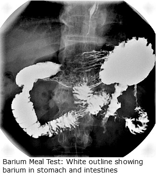
through the material, and it shows up a light color on the film, outlining shadows of the lining of your lower oesophagus, stomach, duodenum and the upper small intestine.
A picture is shown to your right here of one X-ray taken during an upper GI series.
How Is A Barium Meal Test Done?
Barium meal test is easy to do.
- About 200mls or a glass full of high density barium sulphate will be given to you to dink
- You will also be given gas pellets (effervescent mixture) and some citric acid, which is found in lemon juice or orange juice, to help expand the inside of the digestive tract
- Your doctor may then ask you to roll around a bit in order to coat the whole esophagus, stomach and upper small intestines with barium
- There is a muscle between the stomach and the duodenum, and your doctor may decide to give you an injection to help relax that muscle
- X-rays are then taken using a fluoroscope, which can take a very good image of the contrast and the esophagus, stomach and upper small intestine.
The above steps are what is involved in a single contrast barium test.
There is another type of barium swallow test called a double contrast barium meal. In this test air is pumped into your stomach to expand it even more, or the doctor may put some nitrogen or carbon dioxide in your stomach to expand it. The double contrast barium meal is even better for viewing details of the lining of the esophagus, stomach, and duodenum.
Usually a specialist called a radiologist will take the pictures, and he or she will interpret them before sending a printed interpretation to your doctor for your doctor to discuss with you.
What Is This Test Used For?
When you have a barium meal, there are some diseases in the digestive system that can be diagnosed by looking at the pattern of the barium in the esophagus, stomach, and duodenum. These abnormalities include:
- Abnormal constrictions of the
esophagus, stomach, or duodenum like in a condition called achalasia
- Hernias like hiatus hernia
- Peptic ulcers
- Obstructions
- Masses
- Inflammation and erosion in the lining of the esophagus, stomach, or duodenum
- Cancer of the lower esophagus, stomach and duodenum.
Some symptoms, which may encourage your physician to order the barium meal test include the following:
- Difficulty with swallowing
- Vomiting
- Burning pain in the center of the stomach
- Indigestion that is persistent
- A feeling that food is stuck somewhere in the gastrointestinal tract
- If you have swallowed a foreign object
- If you have a vitamin deficiency and your doctor thinks you may be having a problem absorbing the nutrients that should be absorbed by the small intestine.
- If your doctor thinks there is a blockage in your small intestine
- If your have Crohn’s disease
- Unexplained weight loss
- Iron deficiency anemia of unknown cause
How Should I Prepare For A Barium Meal?
Everybody needs adequate preparation before having a barium meal examination. This is to ensure that you get the best result out of the test. It is important that you do the following:
- Do not eat anything at least 6 to 12 hours before the test. This is so as not to have food residue mixing up with the barium and distorting the picture obtainable from the test
- For a day or two before the test, you may have to eat a diet, which is low in fiber. This is not always necessary. Your doctor would advice you, if needed
- Also, in some cases, you may have to take a laxative the night before the test, if your doctor prescribes one, to clean out the intestine
- Take you all your medications as you normally would, unless you are specifically instructed not to
- If you are diabetic, phone in to let the radiologists know, so that you can be given an earlier slot to avoid you having to fast for too long
- Before any test, tell your doctor what
medications you are taking, and if you have allergies to any medications or to
barium or other X-ray contrast materials.
- You must also tell your doctor if you are pregnant or if there is a chance you may be pregnant, because the radiation in the tests, although a small amount, might harm the fetus.
What Can I Expect During The Barium Test?
You will usually have the barium meal test done in the radiology department of your local hospital, but some doctors, specialists in gastrointestinal diseases, may be able to do it in their clinic.
- You will have to remove your clothes and put on a gown, and you will have to take out dentures and take off jewelry.
- During the barium meal test, you will be on you back on an X-ray table, and the table is tilted upright with the X-ray machine positioned in front. There will be a technician who will be with you during the test.
- There is a plain X-ray taken before you drink the barium. This is called a “scout film.” Then you will be asked to take small swallow many times during the multiple X-rays that will be taken.
- The radiologist will be looking at all the
pictures from the X-rays, and he will also be looking through a special camera
called a fluoroscope, which will allow him to see the barium traveling through
you gastrointestinal tract.
- If your doctor has order a test called a “small bowel follow through,” this means the radiologist will take pictures of the entire small intestine, not just the first part.
- The barium meal test takes about 45 minutes at most, but if you have a small bowel follow through test, if may take between two and six hours for the barium to pass through your small intestine.
- Sometimes you may have to return after 24 hours for another X-ray to be certain that the barium has passed into the small intestine and is going into the large intestine.
- After the test, you can usually go home, and you may have to take a laxative or an enema to get all of the barium out of your digestive system. Be sure to drink a lot of fluid after your barium.
In a few days, your doctor will have the results and he will call you to discuss them or perhaps you will be asked to make a clinic appointment if there are any problems that require treatment or further tests.
Are There Any Risks Involved With Having A Barium Test?
The risks involved with barium test examination are very small.
- Barium is not
absorbed into your blood stream, so the chance of allergic
reaction is very very small.
- You may choke or even gag while drinking barium, and possibly, some of the barium may end up inhaled into your lungs. But this is very very rare.
- There may be a chance of
blockage from the barium itself, and if you have an ulcer that has perforated,
there may be some leakage. Usually, if the doctor suspects a perforated ulcer,
he will give you a different type of material to swallow, called Gastrograffin.
- It is also radiopaque. The amount of radioactivity from barium is very small,
but your doctor will warn you that radioactivity of any type may damage cells.
You should keep in mind that we are exposed to small amounts of radioactivity
in the environment all the time. So this should be no problem.
Possible Results And What They Mean
Now you have completed your barium examination. You return to see your doctor to discuss the result. What did the test show, you must wonder. The following are the possible results you can get from doing a barium swallow or meal test:
1. Normal Study
This means exactly what it says. That means there is no abnormality found on the barium study. For many conditions for which barium meal x-ray examination is requested, they can be picked up on the test if there is any abnormality.
Should the radiologist or your doctor still have any doubt as to the reliability of the test for your specific symptom, another test like a CT scan or MRI would be requested.
2. Filling Defects
If there is a report of filling defect seen on your Barium test, it is an abnormal result. It means that your symptoms that led to a barium meal examination being requested might be due to:
- A cancer of the esophagus or stomach or duodenum. this often show up as irregular shouldering with the presence of over hanging edges
- Lymphoma of the stomach
- Polyps
- Trichobezoars (or swallowed hair ball)
- Enlarged spleen could also cause a filling defect (extrinsic)
3. Fold Thickening
Fold thickening in barium meal test refers to the presence of increased tissue shadow at rugae or folds of the inner surface of the stomach. It is seen in conditions like:
- Crohn's disease
- Severe form of gastritis
- Zollinger-Ellison syndrome
Outlet Obstruction
This is a situation seen if the outlet of the stomach or duodenum is narrowed or blocked.
References:
- Chernecky CC, Berger BJ (2008). Laboratory Tests and Diagnostic Procedures, 5th ed. St. Louis: Saunders.
- Fischbach FT, Dunning MB III, eds. (2009). Manual of Laboratory and Diagnostic Tests, 8th ed. Philadelphia: Lippincott Williams and Wilkins.
Barium Meal Examination: Share Your Thoughts Here!
Have you had any experience with barium meal in the past? How did it go? What preparation did you have to make? How do you find the test generally? Would you be happy to do it again if necessary?
Or Are you preparing to have a barium test yourself? Is there something on your mind by way of question you would like to ask?
Post your thoughts here and start a discussion on this topic. We would love to hear from you.
|
Help Keep This Site Going |


