IVU - Intravenous Urogram (Pyelogram)
Last Reviewed: 21st June 2016.
An IVU or intravenous urogram, also referred to as intravenous pyelogram, is a type of test done by injecting a dye into your vein and then taking x-rays of the kidney, ureter and bladder, while the material travels through your urinary tract system. It is used in the identification of many abnormalities, including blockages, presence of kidney or bladder stones and tumors at any point along the urinary tract system.

The kidneys are the upper part of the urinary tract system, and they filter all of the blood as it goes through your body, taking out waste components, balancing electrolytes, and producing urine.
Filtered urine travels down tubes called ureters, which are thin muscular tubes, that is starts from each kidney to the urinary bladder that is located in your pelvis. From the bladder, your urethra, another, short tube, carries the urine outside of your body through an opening called the urethral meatus.
Any disease or blockage along this urinary tract could cause symptoms including abdominal pain. An IVU is one of the tests that could be used to look for what could have gone wrong inside our urinary tract.
Why An IVU?
There are many reasons a doctor might order an intravenous pyelogram.
You may have complained to your doctor about certain symptoms. Those symptoms that might indicate a problem with your urinary tract include:
- Pain the area where your kidneys are located
- Blood in your urine
- An infection in your urine that will not go away with antibiotic treatment
- Difficulty urinating
- High levels of chemicals in your blood that should be excreted by the kidney
- A mass on your kidneys
- A history suggestive of kidney stones
- If you were in an accident or if you had a penetrating injury to the area where your kidneys are located
- If the patient is a child, the doctor may be looking for what is called “vesicoureteral reflux,” which is backflow of urine from the bladder into the ureters
- Acute glomerulonephritis
- Renal Artery stenosis
- Acute Tubular Necrosis (ATN)
What To Expect During An Intravenous Urogram
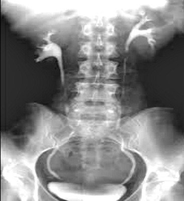
 Picture of an intravenous urogram film highlighting the shadows of the contrast materials (white) emanating from each kidney.
An intravenous urogram is normally done in the radiology suite. The test is done by an X-ray technician and the results are interpreted by a doctor who reads radiologic studies, known as a radiologist.
You will have radiographs taken at various points during the test, so you will have to remember to take off your jewelry and any metal. You will also have to take off your clothing, and will be given a gown or a sheet to cover up. You will have to empty your bladder by urinating just before the test.
You will take the test while you lie on an X-ray table, and the “scout” film, a plain radiograph of your abdomen, will be taken first, to be examined by the radiologist. This is to show that you are in the right position and that there is nothing abnormal that is visible on the scout film.
- You will next receive an injection of contrast dye through one of the veins in your arm, which will be cleaned thoroughly before your injection, using an antiseptic. The contrast dye will travel through your bloodstream until it reaches the kidneys, where it is filtered out, and passes into the ureter from each kidney. It is then drained into the bladder, and is excreted from your body through the urethra.
- During the test, X-ray photographs are taken every few minutes, so the doctor can watch the dye travel through your bloodstream and into the ureters. As each X-ray is taken, it is developed by a technician. If the doctor cannot see what he wants to see, you may have to move around on the table so he can get a complete picture of the urinary tract.
- During the IVU, if the radiologist wants the dye to stay in your stomach for a while, the technician will wrap something around you to compress your abdomen, usually a wide belt with balloons on each side, which will then keep the dye from traveling immediately through the ureters. If you have problems with your abdomen, you will not have to use the belt.
- The X-ray camera will have a beam of X-rays that will be used to show the dye as it takes the pictures. This is called fluoroscopy. The images will be displayed on a video monitor during the test.
The test will take about an hour, and afterwards you should drink a lot of fluids to flush the radioactive contrast material out of your body.
The radiographic pictures do not hurt at all, but the X-ray table may feel uncomfortable after a while. If you have to hold a position, it may also be uncomfortable. When the needle pierces your skin, it may feel like a prick or a sting, and when the contrast dye is injected, you may feel a warm flush.
Some people complain of a salty taste or a metallic taste in their mouths during the test. The compression belt may feel snug or uncomfortable. Some people may experience nausea or lightheadedness after the test.
How To Prepare For An IVU
There are several things that you should tell your doctor before you take the test. These things include:
- If you are or may be pregnant. You should not take the test if you are pregnant, because the radiation could harm the growing fetus.
- Tell the doctor if you have an intrauterine device in place, because the metal may interfere with the test.
- Tell the doctor if you are allergic to iodine, shellfish, or contrast dye. Tell the doctor if you have had an allergic reaction from a bee sting. Tell the doctor if you have had another X-ray study using barium within the past four days. Barium could interfere with the test results.
- Tell your doctor if you use Pepto-Bismol or medications that contain bismuth, which can interfere with the test results.
- If you have kidney problems, or have had them in the past, tell your doctor. Your doctor will take a blood test that will tell him if you can still take the test.
- If you have any concerns about the test, or if you want to know anything about what it means for you, this is the time to ask your doctor.
- Tell the doctor if you have diabetes, and if you take the medicine metformin to control your blood sugar, because it may cause kidney damage.
- Sometimes, your doctor will ask you to take a laxative to clean out your intestines before the test, so that anything in your intestines does not interfere with the test. Your doctor may not want you to eat for 12 hours before the test, to keep your intestines from having food or gas in them.
- If you are breast-feeding, you will have to discard your breast milk for up to two days after the test, because it could have traces of radioactivity in it.
Are There Any Risks To An Intravenous Pyelogram?
- In any test where radiation is used, there may be a very small risk of damage to the cells or tissues of your body. The chance of damage is much smaller than the benefits you can receive from having the test done.
- You could have an allergic reaction, which can be very mild to very severe, but the radiologist will have medicines at hand to control any allergic reaction.
- Some patients who have chronic kidney disease, sickle cell anemia, a type of blood cancer known as multiple myeloma, or pheochromocytoma, could possibly have kidney failure, but that is one reason your doctor will ask you many questions about your health conditions.
What Should I Expect After An Intravenous Urogram?
- Some doctors may have the tests right away, and they will talk to you about the results. If there are any abnormalities, you may need further testing.
- Sometimes the doctor will not get the complete results from the radiologist for one or two days. Then someone from your doctor’s office will contact you to tell you if the test was normal or abnormal.
- In a normal test, the parts of the urinary system are normal in size, position, shape and function. The contrast should flow freely in the normal amount of time, and there will be no blockage.
- In an abnormal test, your doctor may see that your kidneys are the wrong size or shape. Sometimes, the contrast material does not drain in the usual amount of time, or there may be a leak. The kidneys may look scarred. There may be a collection of pus, called an abscess, or a tumor. There may be a blockage because of kidney stones. The doctor can also notice if the prostate gland is too large in a man.
- A picture of part of an IVU is shown below The "pelvis" of the kidneys are see in white towards the top, and the ureters are seen with contrast material outlining them. They empty into the bladder, which is filled with white contrast at the very bottom of the X-ray.
References
- Chernecky CC, Berger BJ (2008). Laboratory Tests and Diagnostic Procedures, 5th ed. St. Louis: Saunders.
- Fischbach FT, Dunning MB III, eds. (2009). Manual of Laboratory and Diagnostic Tests, 8th ed. Philadelphia: Lippincott Williams and Wilkins.


