Kidney Stones
Understanding What Stones In The Kidney Are, How They Are Formed, Who Suffers With Them, And How To Prevent And Treat Them.
Kidney stones are actual stones that form inside the kidney from certain types of salts in our diet or for many other reasons. They could become large enough to cause obstruction and abdominal pain. See signs and symptoms of stones in the kidney and how to prevent and treat them.
Over 350,000 persons are admitted into hospital every year in the United States due to pain and other complications arising from stone in their kidney.
The pain of kidney stone could be so severe that many female sufferers describe it as equal or more severe than childbirth!
It often starts like this. You wake up one morning, or indeed anytime of the day, with a sudden onset severe right or left sided flank pain. The pain is so severe that you clutch onto the side of your abdomen, moving or rolling around to seek a position of comfort. The pain spreads from your upper flank or side of the abdomen towards your groin. It comes in waves - continuous pain but with periods of increased severity. You go pale, feel nauseated and sometimes actually vomits.
You may feel unwell in yourself and in a few sufferers, you may end up passing small amount of urine every few minutes.
What Are Kidney Stone?
Top Tips To Prevent Kidney Stones
- Drink 2 to 6 litres of water daily
- Reduce Your consumption of red meat like beef, pork, or lamb
- Add less salt to food
- Nuts and chocolates are rich in oxalate. Reduce intake.
- Increase your intake of calcium rich food
- Love black peppers? They could be the cause of your kidney stones!
These are real stones formed within the kidney or bladder when the salts or minerals in our urine clump together and grow, instead of being diluted and flushed out of our system. They are also called renal stones, or urolithiasis. They can form from one or a combination of many salts including from:
- Calcium oxalate salts (over 75% of stones are of this type)
- Calcium phosphate
- Uric acid stones
- Cystine
- Ammonium urate
- Sodium urate
- Magnesium ammonium urate
When any of those chemicals normally filtered from our blood accumulate in the kidney, they then "crystallize" depending on a number of factors, and form stones.
These stones could be a few millimeter in size, up to several inches wide and long. Over 80% of stones are less than 5mm in size and are passed away when you urinate without your knowing. Stones as big as a golf ball has been recovered from some sufferers. But, why should some people form stones and not others?
Causes of Renal Stones
The first evidence of stones in the kidney dates back to 4,800 years ago in Egypt. This condition is more common in Europe, North America, and the Middle East. It is thought to be associated with the consumption of food rich in refined carbohydrate and animal protein, with less dietary fibre.
You can develop stones in your kidney or ureter or bladder for a number of reasons. If you are male, you are almost 3 to 4 times more likely to have stones than a woman. If you have it once, you are 5 times more likely to have stones again in your kidney or bladder.
The following are the common causes of kidney stones:
|
|
Of the long list above, the most common causes are chronic dehydration - from poor water drinking habit, dietary choices, and presence of conditions like gout, family history of kidney stones and prolonged use of steroids.
Symptoms
The symptoms of kidney or renal stones actually depends on which part of the kidney or urinary system that the stone is in at the time of symptoms.
Generally, stones in the kidney or bladder do not cause pain or any symptoms, unless they grow too big, causing obstruction or a small chip breaks off and attempts passing down the ureter - the tube that connects the kidney to the bladder, or in the case of a stone in the bladder, if they habour infection or cause irritation and bleeding in the bladder.
The most commonly recognized and described pain due to kidney stones are those of a stone in the ureter.
The following are the typical symptoms of stones in the respective part of the kidney tree or urinary system. They are:
1. Symptoms of Stone Within The Kidney
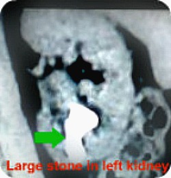
Stones could accumulate and grow within the substance of the kidney, as shown in the picture above. This could go on for many years without any symptoms. When they grow too big, they could cause the following symptoms:
- Continuous nagging dull ache on the side of the abdomen or upper flank. This could be on the right or left side, depending on which of your kidney is affected
- Recurrent urine infection and dull ache in upper flank
- Blood in urine - this is not the only cause of blood in urine
If you have family history of stones in the kidney and suffer with one or a combination of the above symptoms, it would be best you speak with your doctor to explore the possibility of urolithiasis in you.
2. Symptoms of Stone In The Ureter
The ureter is the tube that connects each kidney to the bladder. It passes from the inner side of each kidney through the flank, crossing over the front of the abdomen into the pelvis as it joins the urne bladder on each side.
It is what drains urine from the kidney to be stored in the bladder.
A stone could form inside the kidney and then breaks away, falling down through the right or left ureter. If it gets stuck on its way down, it could cause:
- Sudden onset severe upper right or left side abdominal pain
- Pain is colicky in nature - that is, it comes in waves
- It may spread from the upper side of the abdomen towards the lower abdomen in front or groin
- In some man, it could cause severe pain that spreads to the tip of your penis
- You may find yourself rolling or turning about or indeed pacing about to find a position of comfort
- Retching, nausea and or vomiting
- Fever, if there is associated infection
- Passage of small frequent urine
- There may be visible or only microscopically visible blood in your urine
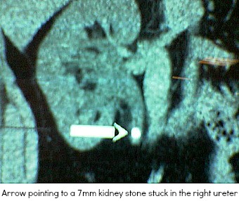
Stones measuring 3 to 7 millimeter in diameter are what typically causes urteric colic. Many times, the stones in the ureter are successfully passed and would cause no other problem. If you have stones in your ureter that his 7 or more millimeter in diameter, they may be more difficult to pass and could cause an obstruction of flow of urine, needing an emergency intervention in the hospital.
3. Symptoms of Stones Within The Bladder
Bladder stones are exactly the same as stones in other part of the urinary system, but this time, they are located within the bladder. They are more common in the elderly or those with restricted movement, or indeed if you are very very prone to stone formation, you could have stones in your kidneys and in the bladder.
Repeated bladder infection is the most common cause and as well as symptoms of bladder stone.
Other symptoms include:
- Dull lower abdominal ache below the naval
- Blood in urine
- Pain on passing urine
- Feeling generally unwell
- Passage of "gravels" in urine
Diagnosis - Tests For Suspected Kidney Stone
If your doctor suspects that you may be having stones in your kidney or indeed your bladder, the following are tests commonly requested and what they expect to show:
- Urine Test. Called urinalysis, you will be asked to pass a sample of urine into a small pot or bottle. This would be tested to see if it shows that there is blood in it. In 80% of cases, there will be blood in the urine - either seen with the "naked eyes" or only microscopically. In the other 20% of cases, there would be no blood. So the absence of blood in your urine does not rule out the possibility of you having a kidney stone. Your urine test may also show white blood cells or leucocytes. In the case of those who suffer with a condition called cystinuria, there would be large collection of crystals of calcium oxalate or urate in their urine.
- Blood Tests. These would include your Complete blood count (also called full blood count), electrolytes and urea, creatinine, uric acid, alkaline phosphate and parathormone levels.
- CT-KUB Scan. Most hospitals now go straight to do a non-contrast CT KUB scan (a CT scan of the Kidneys, Ureters and bladder, done without a dye injected into your blood). They show pictures of kidney stone clearly as can be seen from the pictures above. Those are pictures of stones in the kidney from CT scanning.
- X-Ray / IVU. 9 out of 10 stones can be seen on a plain x-ray of the abdomen. An x-ray of the abdomen is taken and then a dye is injected into your vein and another x-ray is taken at regular intervals intervals of 0, 15, 30 minutes, 2 hours and sometimes 8 hours to study the flow pattern of urine down the ureter and if there is any obstruction. This form of testing is longer done in many places with the coming of CT scanning which is much detailed and requires about the same amount of radiation.
- Ultrasound scan. In children and pregnant women, the decision may be made to do an ultrasound scan of the abdomen. This helps reduce the amount of radiation exposed to. It is also quick and less expensive. The draw back is that sometimes, it may not give enough detail as would a CT scan or MRI.
- MRI. Used in situation where radiation is to be minimized or if extra detail is required.
- Metabolic Screening. This is a special type of urine test done in those in whom a strong suspicion of metabolic disease is made. It involves collecting all the urine they pass over a 24 hour period into a container and then analyzed for the amount of calcium and phosphate in them. They are also tested for the levels of urate, cystine and oxalate in their urine.
Most times, with a combination of two or three of the above testing, a firm diagnosis of kidney stones can be made.
Treatment
Lots of advancements have been made in the treatment of kidney stones. The approach now include:
Adequate Pain Control.
Because most kidney and indeed bladder stones would pass down on their own, the focus for most of these stone is the administration of adequate painkillers. Provided there are no other complications, stones measuring 7mm or less would pass on their own within 24 to 48 hours.
Commonly used pain killers include:
- Diclofenac 100mg suppository inserted into the back passage or rectum in those who have do not have prior kidney problems, do not suffer with asthma and do not have stomach ulcer or on warfarin
- Morphine injection
- Pethidine
Medical Expulsive Therapy
This involves the use of pain killers in conjunction with other medicines to try to get the body to expel the stone. Sometimes, with adequate support, stones up to 10 millimeters can be expelled this way.
Medications used in the medical expulsive therapy include:
- Diclofenac suppository 100mg twice daily into the rectum for 2 to 10 days in combination with
- Steroid tablets and or
- Calcium channel blockers or
- Alpha receptor antagonist.
Active Stone Removal
In those who have stones bigger than 7 millimeter, and are still in a lot of pain despite the administration of good amount of pain killers, they should be offered active stone removal.
Active stone removal is also necessary if:
- Stone obstructing the ureter with associated infection of the kidney
- Someone with a single kidney in whom there is obstruction and or infection
- There is hydronephosis - ballooning of the kidney due to back flow of urine
- Both kidneys are obstructed the same time.
The options for active stone removal available include:
- Extracorporeal Shock Wave Lithotripsy (ESWL). This is a way of treating kidney stones invented in Munich Germany in the 1980s. It employs the use of very high energy sound waves to beak down stones into powder form that is then flushed out in urine. This treatment is done from the outside, without a single cut on your skin. It is a very safe and less painful way of treating stones.
- Percutaneous Nephrolithotomy (PNL). This is the same as an endoscopic or keyhole surgery used to break large difficult to treat stones into small fragments and removed through the skin. usually very big stones that can not be reached or cleared by the ESWL methods are treated using this PNL method.
- Retrograde Intrarenal Surgery (RIRS). This is the use of telescopes to go through the bladder, ureter and then into the kidney to operate and remove stones without any cut on your skin outside. It is a minimally invasive surgery commonly used in children and the obese, and in special cases where extensive surgery is not anticipated.
- Open Surgery. This is rarely used now. It is the traditional way of cutting open the kidney or bladder and the stone extracted under direct vision. A cut of about 30cm is usually made in the flank to achieve this.

Kidney Stones: Have Your Say
Do you or your loved one have kidney stone pain or have a query about kidney or bladder stone treatment? Or do you have a great story or comment on anything related to renal stone? Share it!
|
Help Keep This Site Going |


